


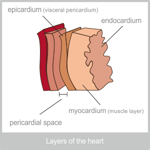
The heart is a cone-shaped, hollow organ that is around 10 cm in length. Your heart is about the same size as your fist. It lies between the lungs slightly off centre to the left, in the cavity under the front ribs. The tip of the cone points downwards and lies roughly 9cm to the left of the mid-line, between the 4th and 5th rib. The heart is made up of three layers of tissue: The pericardium, the myocardium and the endocardium.
 Pericardium
The Pericardium is the outermost layer of the heart and is itself made up of two layers. The outer layer is made of strong fibrous tissue that attaches to the major blood vessels at the top and the diaphragm at the bottom. The inner layer of the pericardium (the serous pericardium) is actually more like a sack. One layer of this sack is attached to the inside of the fibrous layer (the parietal pericardium) and the other side (the visceral pericardium) is attached to the heart muscle itself. The sack is made up of special cells that are able to secrete fluid that fills the tiny space between them. This fluid acts as a lubricant that allows for the smooth movement of the beating heart.
Myocardium
The myocardium is the layer of muscle that allows the heart to beat. It is made up of specialist cardiac muscle cells that are not found anywhere else in the body. These cells link together to form what looks like a sheet of muscle. Each cell is very closely connected to those next to them by special ‘joints’ known as intercalated discs. This arrangement allows the cardiac muscle cells to pass on information very quickly and essentially work as one unit. In other words the contraction of one cell leads to the contraction of the whole heart.
Endocardium
The endocardium is the lining of the heart. It is very smooth, which allows the blood in the heart to flow efficiently.
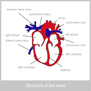 Structure of the heart
The heart is made up of two completely separate halves. The right half pumps de oxygenated blood from the body to the lungs and the left half pumps oxygenated blood from the lungs to the body.
The right side of the heart is the final destination for the largest veins in the body and the left is the beginning of the road for the largest arteries.
Both sides of the heart are further divided into two chambers. The top one is called the atrium and the lower one is called the ventricle.
The muscles around the bottom of the heart – around the ventricle – are thicker than those around the top. This is because the atrium is the chamber that blood is poured into by the body. This then passes through a specialist valve into the ventricle. The ventricle is the section of the heart that actually pumps the blood from the heart. When it contracts the valve snaps shut, stopping the blood from flowing back into the atrium.
On the right side of the heart the blood enters via the two major veins of the body, the Superior and Inferior Vena Cava. The blood from this side of the heart is pumped into the pulmonary artery, and onto the lungs. This is the only artery in the whole body that carries dehydrogenated blood. It needs to be an artery because of the pressure the blood is under. It is also unusual for an artery as it contains a valve at its opening to stop blood flowing back into the heart.
On the left side of the heart the blood comes in from the pulmonary veins. These veins, that come from the lungs are unique because they carry oxygenated blood. The blood is then pumped out of the heart into the aorta. The opening of the aorta is also protected by a valve.
Don’t forget the structure of the cardiac muscle means that the whole heart contracts together. This means that the left and the right ventricle are pumping together. Into the lungs and out to the body simultaneously.
The lud-dub sound of the heart that can be heard through a stethoscope is the sound of the two heart valves closing in sequence. First the valves at the entrance of the ventricle and then the valve at the exit of the heart. When one of the valves does not close properly, some blood leaks back. This leads to a muffled sound that is known as a heart murmur.
The coronary arteries
The heart itself is provided with blood through the coronary arteries. These branch off from the aorta almost as soon as it leaves the heart. They branch out to surround the heart like a net and proved it with the fuel it needs to pump. It is the health of these arteries that is often compromised through smoking. If you think about it when smoke is inhaled into the lungs any contaminants are free to move into the blood. This blood then passes through the pulmonary veins into the heart and then on to the relatively narrow coronary arteries and the aorta. Contaminants from the blood cause the coronary arteries to clog up and lose their elasticity. When we smoke we are quite literally building a cage around our hearts as we congest the fine net of arteries that serve it.
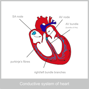 The heart’s conduction system
The heart contains several nodes that are responsible for regulating the heart beats. They are made of specialised neuromuscular tissue.
Sinuatrial node (SA node)
The SA node, is on the right side of the heart near the opening of the Superior Vena Cava.
The atrioventricular node (AV node)
The AV node is between the two halves of the heart. It is usually stimulated by the impulse that comes from the SA node,
The SA node initiates faster beats than the AV node, so it is usually the SA node that works as ‘the pacemaker’. However, the AV node it is capable of initiating beats, but it does so more slowly than the SA node.
The AV node, situated as it is between the two halves of the heart branches down through the separating wall and all around the base of the heart. When it receives and impulse from the SA node it sends a message to the muscles around the base of the cone. This begins the wave of contraction that results in the pump of the heart.
This mechanism allows the heart to function independently, but our heart beats can also be controlled by our brains through the autonomic nervous system. The cardiac centre of the brain is in the medulla oblongata. The autonomic nervous system consists of two aspects that work together to keep us in balance. These are the parasympathetic and the sympathetic systems. In relation to the heart, parasympathetic stimulation, reduce the rate of impulses and therefore, reduces the speed and force of heart beats. Sympathetic stimulation, on the other hand, increases the rate of impulses and therefore increases the rate and force of the heart beat.
Factors that can affect the rate of the heart include, exercise, temperature, emotional arousal, age and gender. (Children and females have relatively higher heart rates)